Macular degeneration is the leading cause of vision loss in the United States.
If you have just been diagnosed with macular degeneration, the thought of losing vision may take your breath away, and with good reason.
Macular degeneration is the leading cause of vision loss and blindness for adults over age 65. More than 20 million Americans already suffer from macular degeneration and that number is only expected to increase as baby boomers age according to the National Institute of Health.
While there is no cure for macular degeneration, there are treatments available to slow the progression of the disease and preserve vision. To catch signs of the disease early, see your eye doctor once a year, especially after age sixty-five.
Macular degeneration begins with damage of the light-sensitive tissue at the back of the eye called the retina. Left untreated, macular degeneration causes devastating blind spots and loss of central vision. The greatest risk factor for macular degeneration is aging, rising from two percent in your forties to nearly 50 percent once you reach the age of eighty.
Macular degeneration occurs specifically when cells within the macula deteriorate. That’s the small area of the retina responsible for sharp central vision used for focusing on things directly in front of you. There are two types of macular degeneration: dry and wet. Dry macular degeneration is the most common type, accounting for nearly 90 percent of all cases. Wet macular degeneration is less common but more severe and often leads to rapid vision loss.
There are several factors that increase the risk for macular degeneration including age, family history, smoking, obesity, and exposure to ultraviolet light. While you can’t change your age or family history, not smoking and maintaining a healthy weight can significantly reduce your risk in addition to wearing sunglasses that limit exposure to harmful UV rays.
Since there is no pain associated with macular degeneration, you may not notice problems with your vision until the disease has progressed significantly. The only way to know if you have macular degeneration, or any other type of eye disease, is from a comprehensive eye exam with your eye doctor.
If you think you may be at risk for macular degeneration, schedule regular eye exams to monitor for subtle changes and to begin early treatment if necessary.
What is Macular Degeneration
Macular degeneration starts at the back of the eye in the retina. The retina is made up of delicate nerve tissue, just like the brain. Once light reaches the back of the eye, visual images are transferred into electric signals and sent to the brain along the optic nerve. In the center of the retinal nerve tissue is an area called the macula.
The macula is a small spot responsible for big impact with your vision. It captures visual signals and sharpens them into the clear, central vision used for reading, driving and seeing fine detail. When the macula is damaged it causes missing and blurry areas in the middle of your vision.
There are two types of macular degeneration: dry and wet.
Dry macular degeneration is the more common type, accounting for nearly 90 percent of all cases. Typically, dry macular develops slowly and painlessly causing a gradual deterioration in vision over time.
Wet macular degeneration is less common but more serious, accounting for about 10 percent of all cases. It often develops suddenly and can cause rapid vision loss.
Macular degeneration causes vision loss in several different ways.
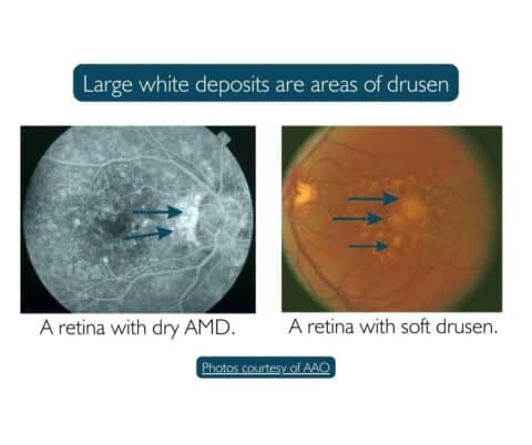
Dry macular degeneration causes vision loss when waste products called drusen build up in the macula, causing it to thin and atrophy. Drusen are small and large yellow deposits under the retina made of fats and proteins. The appearance of drusen does not necessarily lead to eye disease but increases the risk. Large particles of drusen are more often found in dry macular degeneration.
Fundus photos of retina with drusen in dry macular degeneration
Vision damage from drusen is usually slow and painless. It can go undetected for extended periods until it has caused some degree of vision loss. Your eye doctor can detect the appearance of drusen with an eye exam.
In its early stages, dry macular degeneration shows no visible changes to the retina, and you may not experience any symptoms.
As dry macular degeneration progresses, significant thinning and atrophy of the retina is detected. At this point, central vision becomes blurred and distorted and sharp details are hard to see. Faces may become unrecognizable and activities that rely on clear central vision like driving and reading become difficult or impossible.
There is no treatment for dry AMD, but it can advance to the wet form at any time which makes it imperative to detect early.
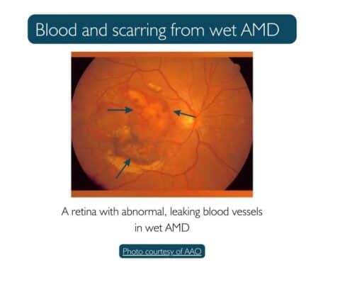
Wet macular is less common than dry, but causes faster vision loss. Wet macular degeneration occurs when unstable blood vessels grow under the retina. These vessels are very fragile and unhealthy. They leak blood and fluid causing the retina to swell resulting in rapid vision loss. Wet AMD often develops suddenly and leads to severe vision loss within days or weeks.
Fundus photo of leaking blood vessels and scarring from wet macular degeneration
Two Types of Wet AMD
Neovascular AMD (also called “exudative” or “disciform”) occurs when abnormal blood vessels grow underneath the retina. Neovascular AMD is responsible for 90 percent of the severe vision loss in wet macular degeneration.
Geographic atrophy refers to a large area of cell death in retina. Geographic atrophy typically progresses slowly and can lead to significant central vision loss over time. This form is responsible for 10 percent of wet AMD cases.
Both forms of wet and dry macular degeneration cause similar symptoms:
Blurry or distorted vision
Difficulty reading or seeing fine details
A dark or empty area in the center of your vision
Colors that seem dull and less bright
How Do I Know if I Have Macular Degeneration?
Macular degeneration is diagnosed after a comprehensive eye exam with your eye doctor that includes a visual acuity test to measure how well you see at different distances, along with a detailed look at your retina. If your eye doctor suspects macular degeneration, additional tests called optical coherence tomography and fluorescein angiography may be ordered.
Optical Coherence Tomography (OCT)
OCT is a non-invasive test that uses light waves to take cross-sectional pictures of the retina. This imaging shows in minute detail any thinning or thickening of the retina caused by macular degeneration. The images are used as a baseline of retinal health and to monitor for significant changes in follow-up visits.
Fluorescein Angiography (FA)
FA is a test that uses injected dye to highlight any abnormal blood vessels in the eye and the choroid tissue underneath. A special laser lights up the dye and shows areas of new blood vessel growth underneath the retina, a common sign of wet macular degeneration.
How is Macular Degeneration Treated
Unfortunately, there is currently no cure for macular degeneration. But there are highly effective treatments that can help slow the progression and preserve vision, especially when started early. These include nutritional supplements, medication injected into the eye, laser surgery, and photodynamic therapy.
There is no one-size-fits-all treatment for macular degeneration. The best course of treatment depends on the individual, their medical history, and the existence of other eye conditions. The type of treatment is dependent on the timeliness of diagnosis and the severity of vision loss.
First line treatment for wet macular degeneration requires monthly or bimonthly injections of an anti-vascular endothelial growth factor medication to inhibit growth of unstable blood vessels in the eye. This anti-VEGF treatment is effective in slowing the progression of wet macular degeneration and preventing further vision loss. Intravitreal injections have also been shown to improve vision for many patients with wet macular degeneration.
Anti-VEGF medication requires ongoing treatment to prevent the recurrence of blood vessel growth and resulting vision loss. The frequency of injection depends on a patient’s response to the medication. Follow-up appointments are typically every 4 – 6 weeks with a retina specialist. For patients receiving these sight saving injections it is important to maintain regularly scheduled appointments. Missed treatments can turn back the clock and potentially worsen vision.
Laser treatments and photodynamic therapy are also used for patients with wet macular degeneration. With photodynamic therapy, a light-sensitive drug is injected into the bloodstream to mark abnormal blood vessels. A laser lights up the abnormal cells and destroys them to stop bleeding underneath the retina.
Laser and photodynamic therapies are not used as often as anti-VEGF treatment or with rapid vision loss from wet macular degeneration. It can be used to augment VEGF treatment or when vision loss as progressed more slowly in wet AMD.
Risk Factors of Macular Degeneration
There is no specific cause of macular degeneration, but there are several risk factors, some within your control and some not.
Uncontrollable risk factors:
Age: Like many health conditions, macular degeneration is more common with age, especially for those over 65.
Family History: You are at increased risk for the disease if you have a parent or grandparent with macular degeneration,
Race:Caucasians are more likely to develop macular degeneration than African Americans or Hispanics/Latinos.
Controllable risk factors:
Smoking: Smoking is the greatest controllable risk factor for macular degeneration. Smoking doubles the risk of developing macular degeneration (along with cancer, heart, and lung disease!) If you don’t smoke, please don’t start. If you do smoke, check out the many free resources to help you quit.
Diet: Obesity and high cholesterol raise the risk of developing macular degeneration due to elevated cholesterol deposits in the small blood vessels.
Diabetes: Diabetics Type I and II are especially at risk for developing macular degeneration along with a condition called retinopathy. Unstable blood sugar levels can cause small blood vessels in the eye to break and leak.
What to do if you are worried about have Macular Degeneration.
Early treatment can slow progression of eye disease and prevent vision loss.
“If you are diagnosed with macular degeneration, it is important to see your eye doctor regularly for follow-up. Regular eye exams are always your first line of defense to detect the early signs of eye disease and begin treatment,” says Dr. S Rana, MD, board-certified ophthalmologist at St Lucie Eye.
With early detection and treatment, you can help slow the progression of macular degeneration and preserve your vision.
During a comprehensive eye exam, your eye doctor will check for signs of macular degeneration and other eye conditions that may have similar symptoms. If you have risk factors for macular degeneration, such as a family history of the disease, your doctor may recommend more frequent visits.
There is no surefire way to prevent vision loss from macular degeneration, but there are steps you can take to prevent or slow its progression.Early detection and treatment are the most important steps to prevent vision loss from macular degeneration. Other healthy habits include not smoking, eating a varied diet with lean proteins, fruits and vegetables, exercising regularly, and wearing sunglasses.
If you are diagnosed with macular degeneration, your eye doctor will discuss the condition in clear, compassionate language for you to understand. You’ll be informed of helpful treatment options to slow progression and preserve your vision. Should you need advanced treatment, a follow-up appointment can be made with Dr. Kevin Kelly, retina specialist at St Lucie Eye in Fort Pierce or Port St Lucie.